Staged groin flap for a degloved dorsum of the hand
A degloving injury of the hand with tendon exposed and no local tissue to close it, resurfaced with a pedicled groin flap. The hand was carried on the abdominal wall for three weeks before the pedicle was divided.
Flap reconstruction · Hand · 25–30 · Male · 6 min read
- Mechanism
- Degloving injury of the hand
- Anaesthesia
- General
- Stages
- Two stages
Presentation
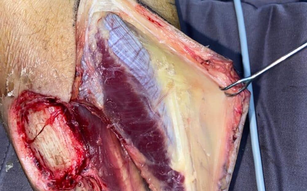
The patient presented with a degloving injury of the hand which had removed the skin and subcutaneous tissue from the dorsum, leaving extensor tendon exposed over a wide area. Exposed tendon does not accept a skin graft: without its paratenon a graft will not take, and a tendon left uncovered dries, adheres and is eventually lost. The problem was therefore not closure but cover — bringing in tissue with its own blood supply.
Full-thickness soft-tissue loss across the dorsum of the hand with extensor tendon visible in the base of the wound and no paratenon remaining over it. The surrounding hand skin was too tight and too damaged to provide a local flap. The groin was unburned and uninjured, with the superficial circumflex iliac territory intact and adequate laxity to close the donor site directly.
Procedure
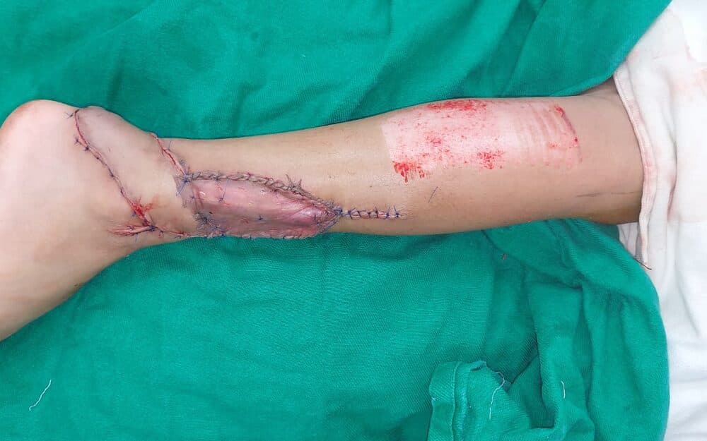
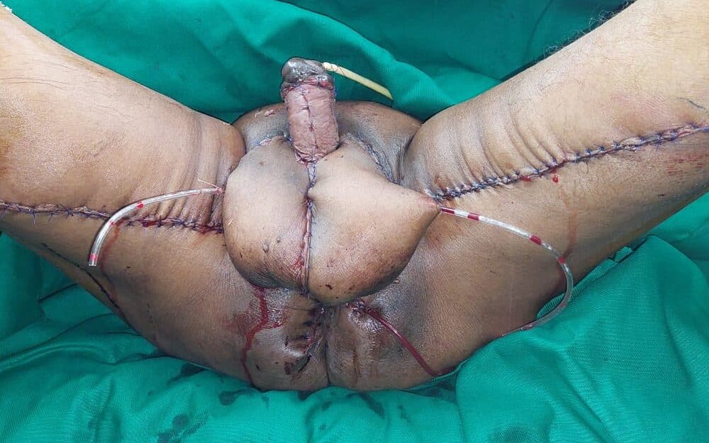
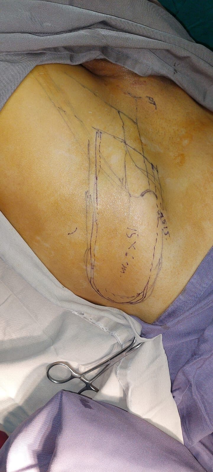
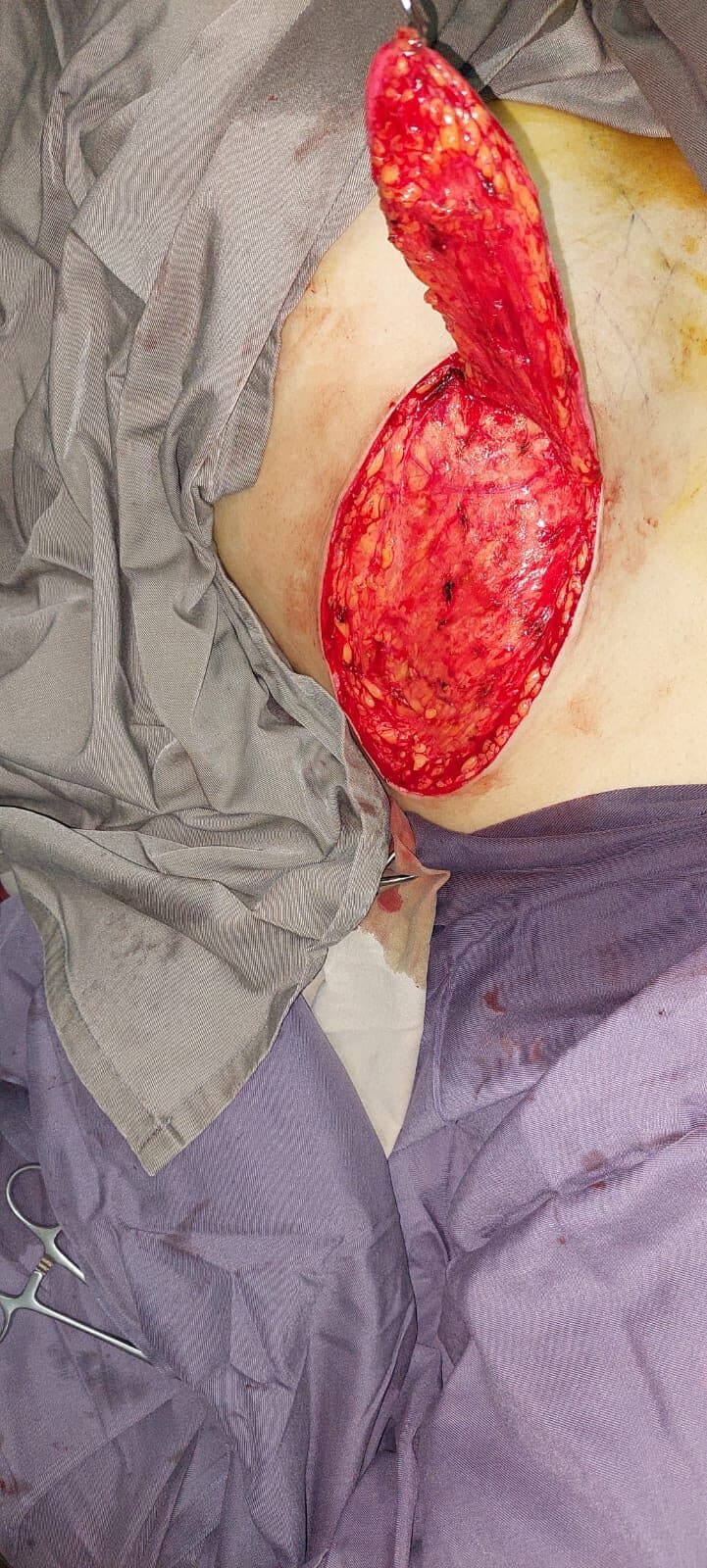
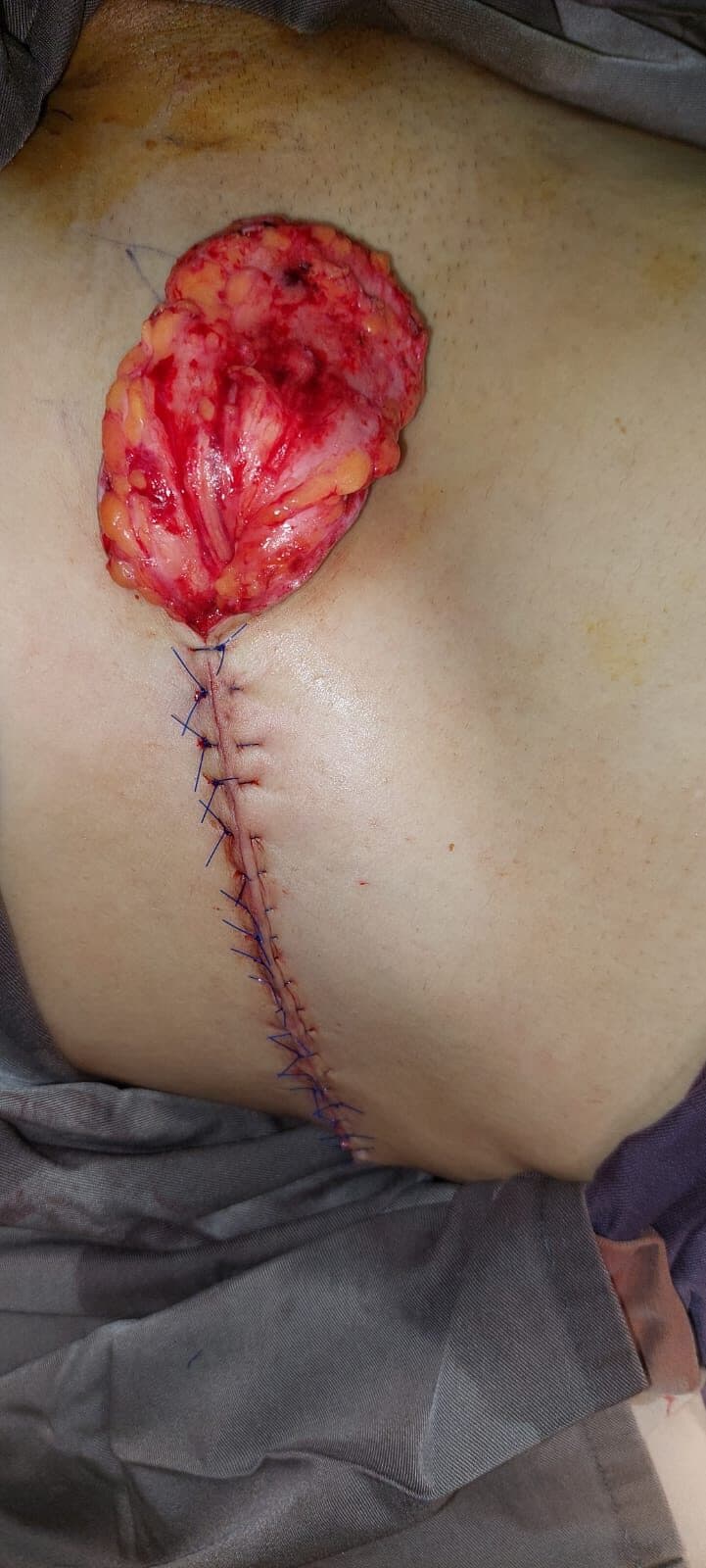
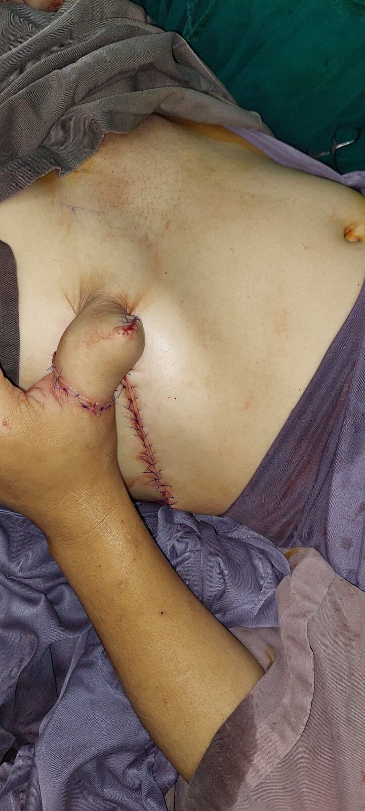
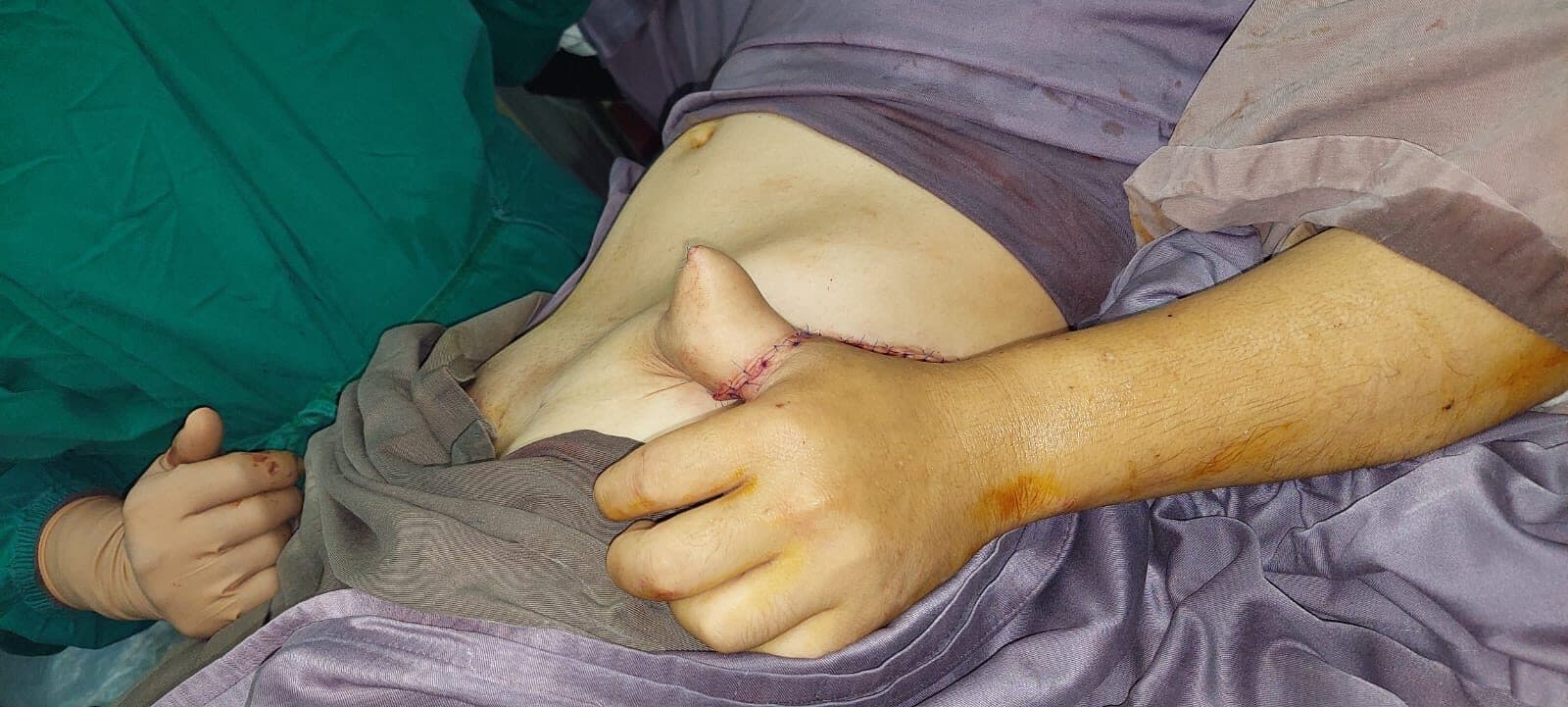
The flap was designed on the groin, marked to the size of the defect and raised on its pedicle, with the donor site planned so that it could be closed directly rather than grafted. The hand was then inset into the flap and the wound edges sutured, so that the limb was carried attached to the abdominal wall while the flap took up a blood supply from its new bed. This is the part patients find hardest — the hand is fixed to the trunk for three weeks and the shoulder and elbow stiffen if they are not moved deliberately. At the second stage the pedicle was divided and the flap inset was completed around the dorsum.
- Day 0Assessment; wound débrided and dressed
- Stage 1Groin flap raised and the hand inset into the flap pocket
- Post-opLimb positioned and supported; flap perfusion monitored
- Week 3Pedicle divided and the flap inset completed
Outcome
The flap survived both stages and provided durable, vascularised cover over the exposed tendon, which is what makes later tendon surgery possible if it is needed. Detailed follow-up, range of motion and return to work are placeholder and await the surgeon’s records.
Clinical images are blurred until you choose to view them. Each is labelled with its timepoint.
Placeholder — to be replaced with the patient’s own words, given with consent.
Surgeon's note
A pedicled groin flap is not the most elegant answer to a dorsal hand defect, and in a unit with reliable microsurgical support a free flap avoids three weeks of the hand strapped to the abdomen. What the groin flap has in its favour is that it works without a microscope, without a long anaesthetic, and without a vascular anastomosis that can fail overnight — which in this setting is often the difference between cover and no cover. The cost is stiffness, and the cost is paid by whoever supervises the physiotherapy afterwards.
Consent
CONSENT PENDING — this photograph is published for build purposes only and must not go live until written consent is on file.
These cases are published for clinical and educational purposes. Every patient, injury and recovery is different: the results shown are individual outcomes and are not a prediction or guarantee of the result of any other person’s treatment. All surgery carries risk, which is discussed in full during consultation.