Reverse sural artery flap for an exposed Achilles tendon
A wound over the Achilles region with tendon exposed, resurfaced with a reverse-flow sural artery flap raised from the calf and turned down on its pedicle.
Flap reconstruction · Lower limb · Adult · 6 min read
- Mechanism
- Placeholder — mechanism to be supplied
- Anaesthesia
- General
- Stages
- Single stage
Presentation
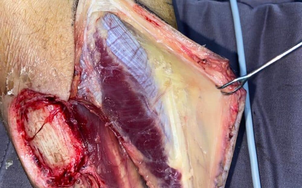
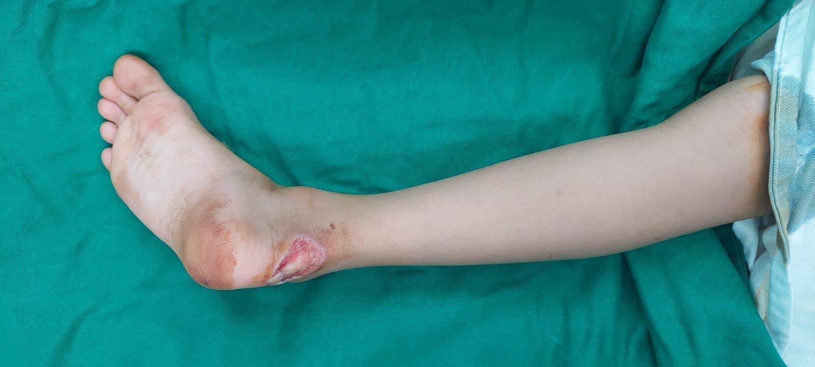
The patient presented with a soft-tissue defect over the posterior ankle with the Achilles tendon exposed at its base. The skin over this region is thin, has little underlying tissue to recruit, and sits over a tendon that must glide — the three things that together make it one of the least forgiving sites in the lower limb to close.
Full-thickness soft-tissue defect over the Achilles region with tendon visible and no local tissue available for direct closure or a local flap. The calf skin proximal to the defect was intact and the sural territory uninjured, allowing a reverse-flow flap to be designed on the posterior calf with its pivot point above the lateral malleolus.
Procedure
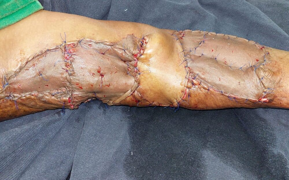
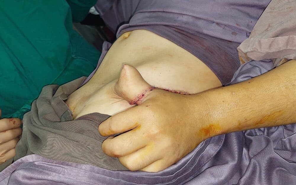
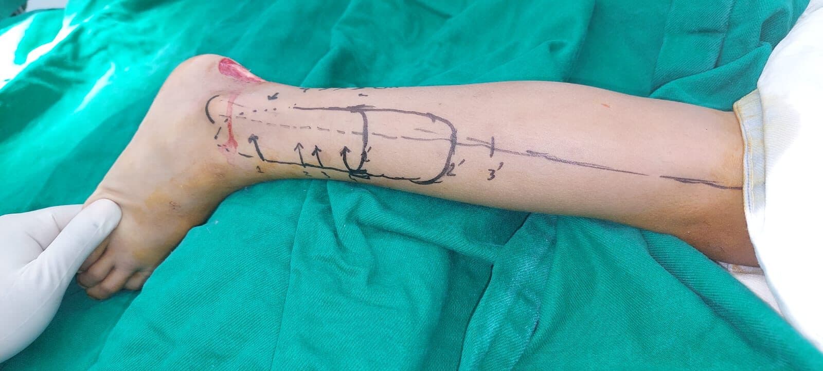
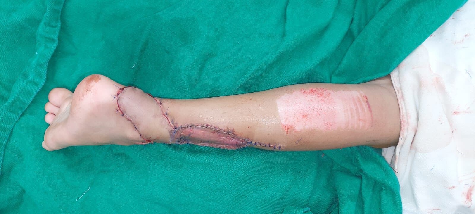
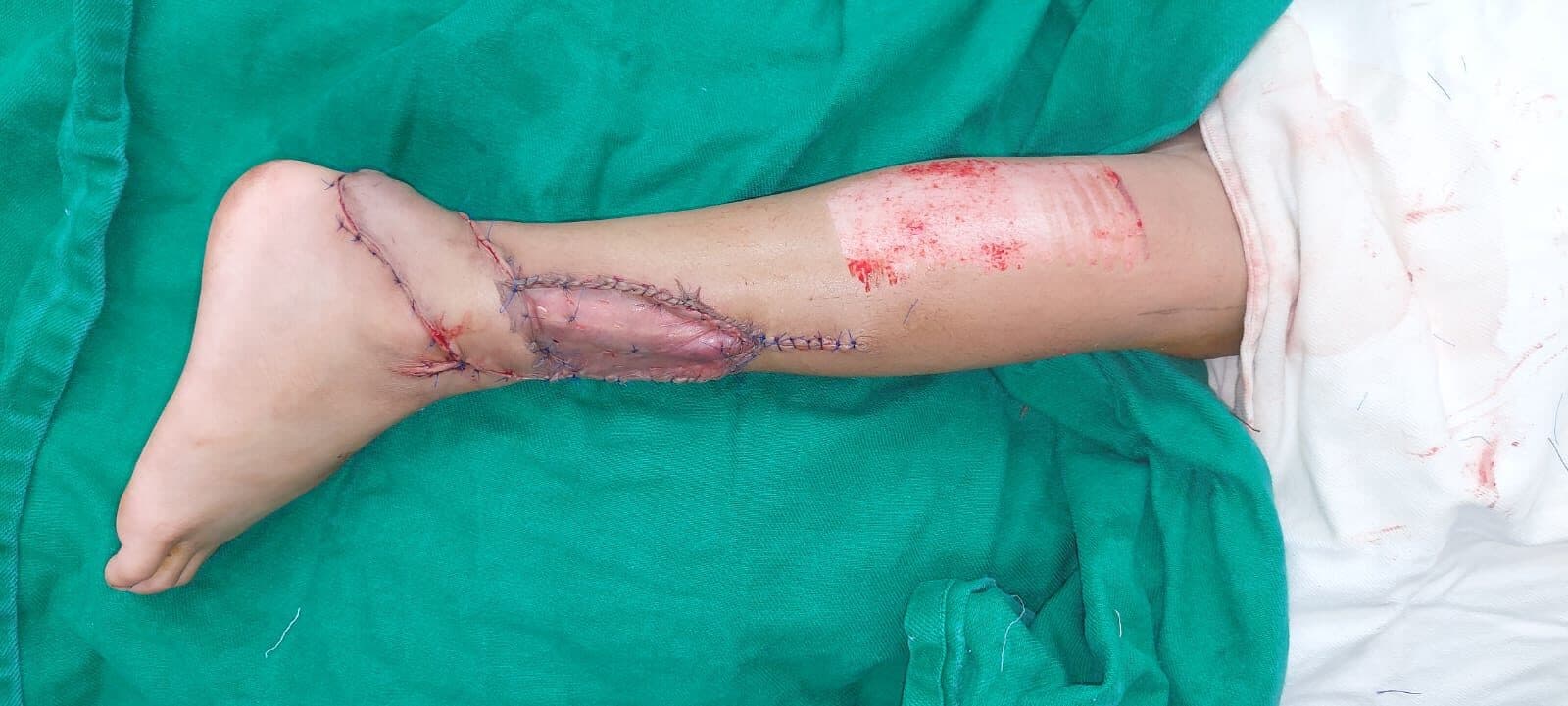
The flap was marked on the posterior calf with its axis over the sural nerve and short saphenous vein, and its pivot point sited a fixed distance above the lateral malleolus — that distance is what protects the perforators the reverse flow depends on, and moving it distally to gain reach is how these flaps are lost. The flap was raised with a wide adipofascial pedicle rather than a skeletonised one, turned down through 180 degrees and inset over the exposed tendon without tension. The donor site was closed and the ankle supported.
- AssessmentDefect assessed, tendon exposure confirmed, flap planned
- OperationReverse sural flap marked, raised and turned down to the defect
- InsetFlap inset over the tendon; donor site closed
Outcome
The flap reached the defect and was inset over the exposed tendon with vascularised cover restored. Flap survival to full maturity, donor-site healing and ankle function are placeholder pending the surgeon’s follow-up.
Clinical images are blurred until you choose to view them. Each is labelled with its timepoint.
Placeholder — to be replaced with the patient’s own words, given with consent.
Surgeon's note
The reverse sural flap has a reputation for venous congestion, and most of that reputation is earned at the moment of design rather than in the dissection. Keeping the pivot high, carrying a broad adipofascial pedicle rather than a narrow one, and refusing to twist it more than the arc genuinely needs — those decisions are made before the knife. Over the Achilles it earns its place because the alternative in most units is a free flap, and this reaches without a microscope.
Consent
CONSENT PENDING — this photograph is published for build purposes only and must not go live until written consent is on file.
These cases are published for clinical and educational purposes. Every patient, injury and recovery is different: the results shown are individual outcomes and are not a prediction or guarantee of the result of any other person’s treatment. All surgery carries risk, which is discussed in full during consultation.