Local perforator flap for a chronic ulcer over the tibia
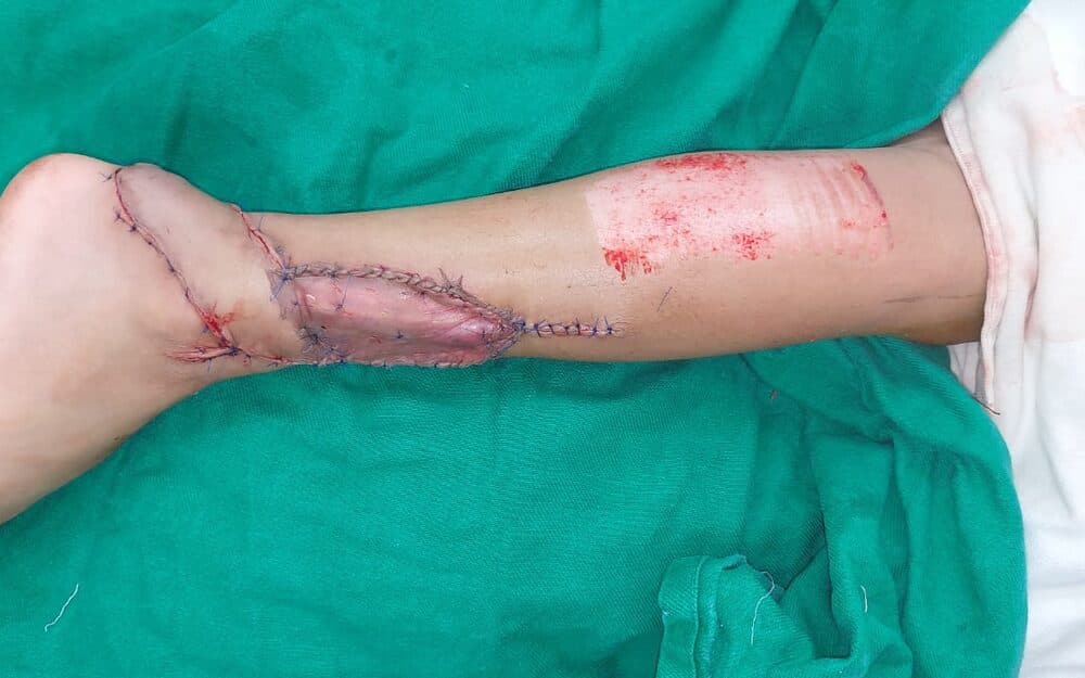
A long-standing ulcer over the subcutaneous border of the tibia, excised down to healthy tissue and closed with a local perforator-based flap raised from the adjacent calf.
Flap reconstruction · Lower limb · Adult · 6 min read
- Mechanism
- Chronic ulceration over the tibia
- Anaesthesia
- General
- Stages
- Single stage
Presentation
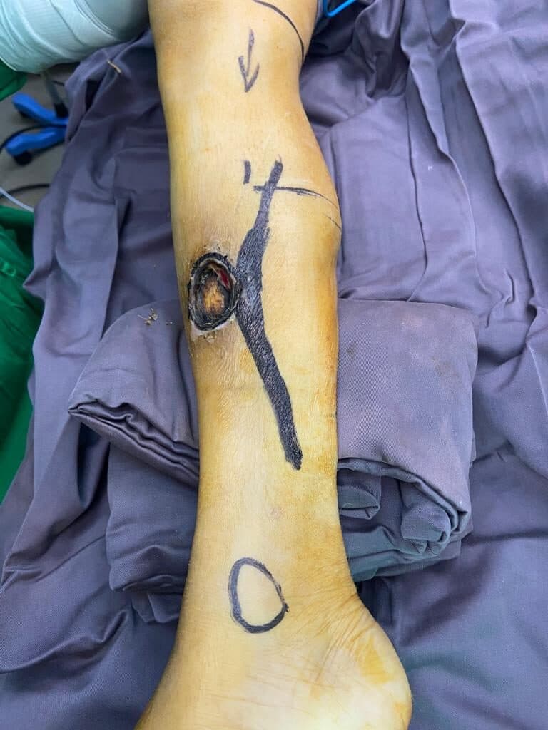
The patient presented with a chronic ulcer over the subcutaneous border of the tibia which had failed to heal with dressings. Over the shin there is almost nothing between skin and bone, so an ulcer here reaches cortex early and then stops making progress: bare cortical bone has no blood supply of its own to granulate from, and no dressing regimen changes that.
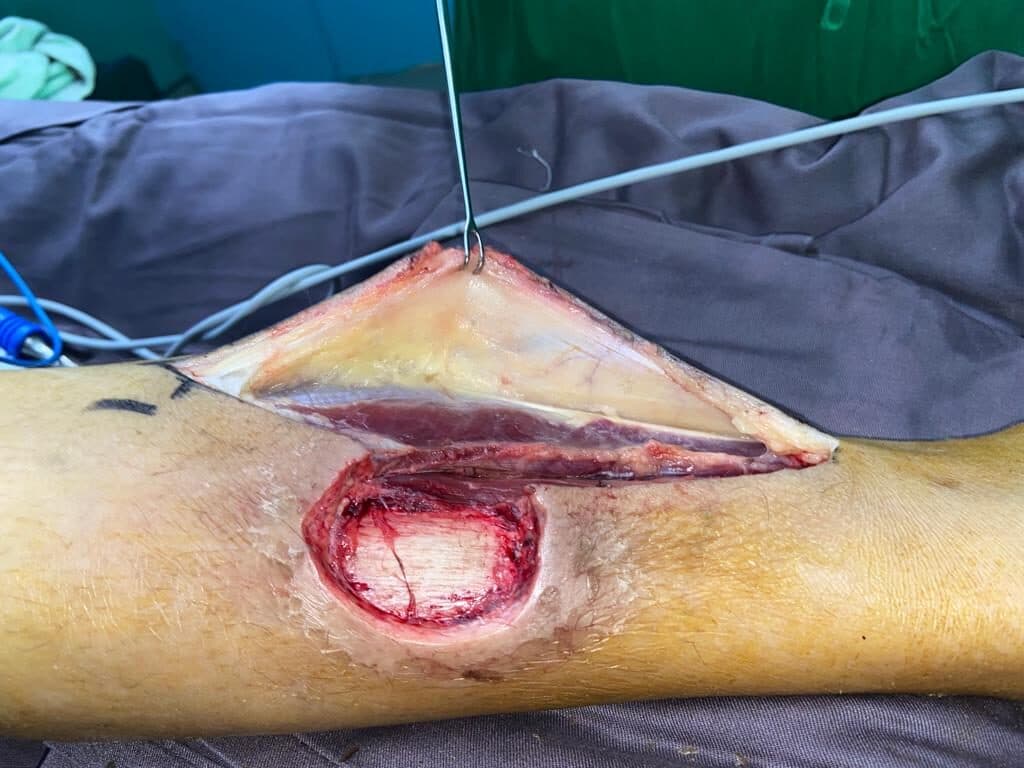
A chronic ulcer over the anteromedial tibia with a fibrotic margin and bone at the base of the wound. The surrounding skin was scarred but the adjacent calf was uninvolved, with a perforator identified proximal to the defect on which a local flap could be based.
Procedure
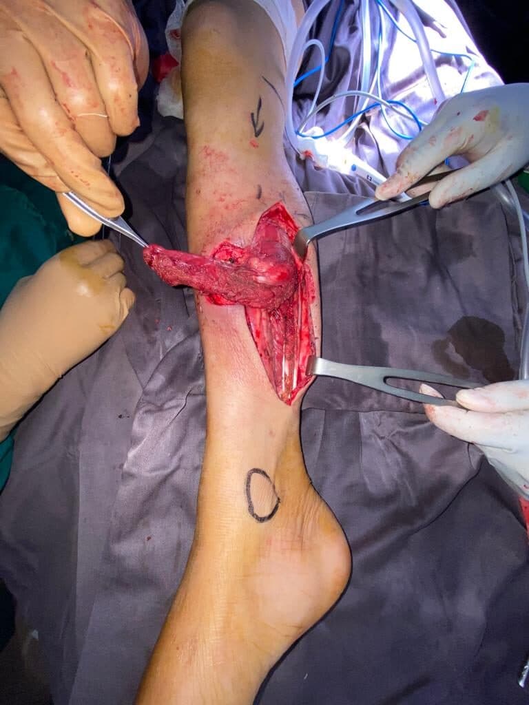
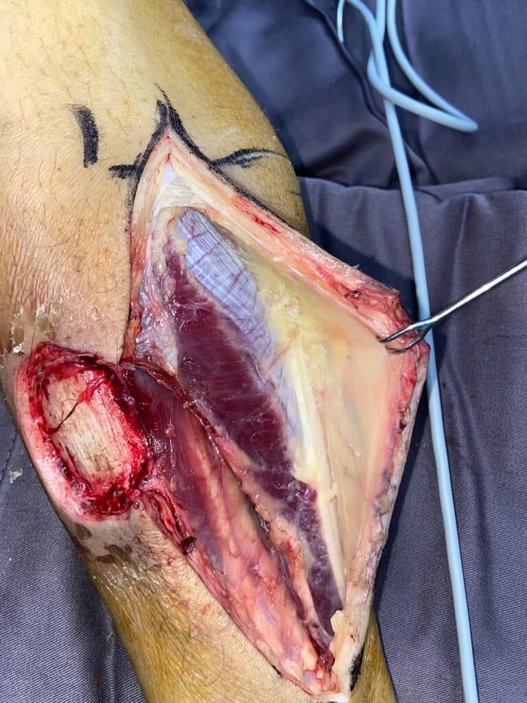
The ulcer was excised in full, including its fibrotic margin — leaving the chronic edge behind is why these recur, and the excision has to be planned as though it were a tumour margin rather than a débridement. The perforator was identified and a local fasciocutaneous flap raised on it, dissected far enough to give the arc of rotation needed without kinking the vessel at the pivot. The flap was transposed into the defect and inset, and the donor area closed.
- AssessmentUlcer assessed; perforator located and flap planned
- ExcisionUlcer and fibrotic margin excised down to healthy tissue
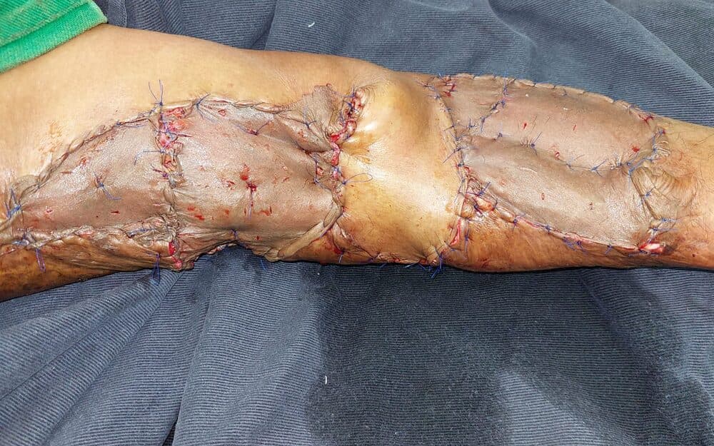
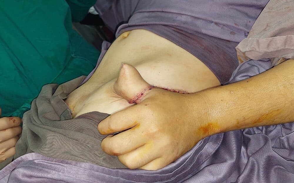
- ReconstructionPerforator flap raised and transposed into the defect
Outcome
The ulcer was excised and the resulting defect closed in a single stage with vascularised local tissue rather than a graft on bone. Healing and recurrence at follow-up are placeholder and await the surgeon’s records.
Clinical images are blurred until you choose to view them. Each is labelled with its timepoint.
Placeholder — to be replaced with the patient’s own words, given with consent.
Surgeon's note
Two things decide a tibial ulcer. The first is that the excision must be complete: a chronic ulcer has a fibrotic, poorly vascularised margin, and a flap sutured onto that margin is a flap sutured onto the reason the wound never healed. The second is that bare cortex needs vascularised tissue, not a graft — grafting onto bone in this position fails, then costs the patient another operation with a worse starting point. A local perforator flap achieves both without committing to free tissue transfer.
Consent
CONSENT PENDING — this photograph is published for build purposes only and must not go live until written consent is on file.
These cases are published for clinical and educational purposes. Every patient, injury and recovery is different: the results shown are individual outcomes and are not a prediction or guarantee of the result of any other person’s treatment. All surgery carries risk, which is discussed in full during consultation.